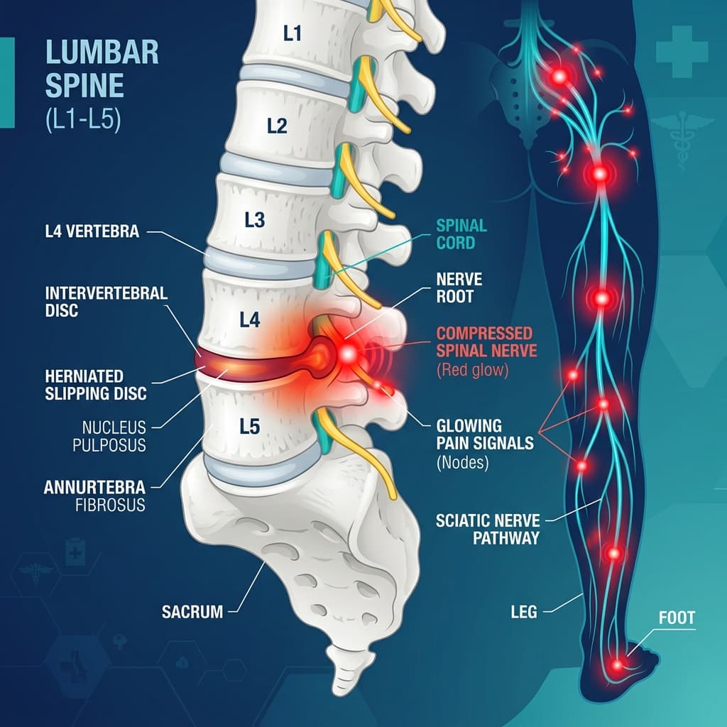
An intervertebral disc is a specialized shock-absorbing cushion positioned between the bony vertebrae of the spinal column. Each disc consists of a tough, fibrous outer ring (annulus fibrosus) enclosing a soft, gel-like inner core (nucleus pulposus). A 'slip disc'—medically termed a Herniated Intervertebral Disc (HIVD) or Disc Prolapse—occurs when tears or micro-cracks in the outer annulus allow the inner gel to bulge or leak outward into the spinal canal.
In India, sedentary desk jobs, improper lifting techniques, long commutes, and poor core strength have made slip disc one of the leading causes of chronic back pain and nerve radiculopathy. Recognizing symptoms early prevents chronic nerve compression, muscle atrophy, and long-term functional disability.
Core Symptoms of a Lumbar Slip Disc (Lower Back & Sciatica)
Approximately 90% of slip discs occur in the lumbar spine—most frequently at the **L4–L5** and **L5–S1** vertebral levels. When a herniated disc presses against lumbar spinal nerve roots, it causes a characteristic cluster of symptoms:
1. Sharp Radiating Sciatica Pain
Sciatica is the hallmark symptom of a lumbar slip disc. Rather than remaining in the back, the pain travels along the path of the sciatic nerve—shooting from the lower back through the buttock, down the back or side of the thigh, into the calf, and extending into the foot or toes. Patients describe it as an electric shock, burning ache, or sharp stabbing pain that makes walking or standing difficult.
2. Numbness & Tingling (Paresthesia)
Mechanical compression of nerve fibers disrupts normal sensory signaling. Patients frequently report 'pins and needles', crawling sensations, or complete loss of feeling in specific skin zones (dermatomes)—such as the top of the foot, big toe webspace (L5 root), or outer edge of the heel (S1 root).
3. Muscle Weakness & Foot Drop
Sustained nerve root compression impairs motor signals to leg and foot muscles. L5 nerve root compression causes weakness in pulling the foot upward (ankle dorsiflexion), leading to 'Foot Drop' where the toes drag while walking. S1 root compression makes standing or pushing off on tiptoes difficult.
4. Pain Aggravation on Bending, Sitting, or Coughing
Intra-discal pressure increases dramatically when sitting or bending forward. Activities like sitting in a car, tying shoelaces, or straining during coughing and sneezing push the gel nucleus harder against the inflamed nerve root, triggering sharp bursts of pain.
5. Paraspinal Muscle Spasms & Spinal Stiffness
The back muscles surrounding the injured disc undergo intense involuntary spasms as a protective protective mechanism to immobilize the painful spine segment. This creates severe stiffness and forces the body to tilt sideways (sciatic scoliosis).
Cervical Slip Disc Symptoms (Neck & Upper Limb)
When disc herniation occurs in the cervical spine (neck)—most commonly at **C5–C6** or **C6–C7**—the symptoms affect the neck, shoulders, and upper limbs:
Key symptoms of a cervical slip disc:
- Cervical Radiculopathy: Sharp shooting pain radiating from the neck down the top of the shoulder, bicep/tricep, and into the arm.
- Hand & Finger Numbness: Tingling or numbness localized to the thumb and index finger (C6 root) or middle finger (C7 root).
- Loss of Fine Motor Grip Strength: Difficulty buttoning shirts, holding a pen, opening jar lids, or dropping objects frequently.
- Neck Stiffness & Suboccipital Headaches: Restlicted neck rotation accompanied by aching headaches starting at the base of the skull.
Interactive Slip Disc Explorer, Nerve Root Mapping & Self-Checker
Use our interactive diagnostic tool below to explore disc herniation severity stages, map radiating pain pathways, and evaluate your spine health symptoms.
Slip Disc Pathology, Nerve Mapping & Assessment Tool
Use these interactive diagnostic tools to understand disc herniation stages, nerve root radiation pathways, and spine health triage.
4 Stages of Intervertebral Disc Herniation
Disc Bulge (Protrusion)
The inner gel (nucleus pulposus) presses outward, causing the disc wall to bulge into the spinal canal without tearing the outer ring.
Disc Prolapse (Herniation)
The gel tears through inner fibers of the annulus wall, creating a distinct bulge that directly compresses adjacent spinal nerve roots.
Disc Extrusion
The gel-like nucleus breaks completely through the outer wall of the annulus fibrosus, extending beyond the intervertebral space.
Disc Sequestration (Free Fragment)
A fragment of the gel nucleus breaks free entirely from the main disc body and lodges in the spinal canal space.
Disc Prolapse (Herniation)
Triggers classic sharp radiating Sciatica down the leg or radiculopathy into the arm, accompanied by numbness.
Short-term anti-inflammatory medication, targeted spinal physiotherapy, lumbar traction, or epidural steroid injections.
Spinal Nerve Roots & Pain Radiation Paths
L4 – L5 Level (L5 Nerve Root Compression)
Radiates from lower back through side of hip, down outer side of thigh and calf to the top of foot.
Numbness or tingling over the top of foot and big toe webspace.
Difficulty lifting foot upward (Foot Drop / ankle dorsiflexion weakness).
Bending forward, lifting heavy weights, sitting for long hours, coughing/sneezing.
Slip Disc & Sciatica Self-Assessment Screener
Select symptoms you have been experiencing for a tailored clinical recommendation.
The 4 Clinical Stages of Disc Herniation
Spine specialists categorize disc herniation into four distinct pathological stages based on MRI imaging findings:
Pathological Stages of Intervertebral Disc Herniation
| Herniation Stage | Anatomical Description | Annulus Integrity | Clinical Severity & Treatment |
|---|---|---|---|
| Stage 1: Protrusion (Disc Bulge) | Nucleus presses outward against annulus ring | Annulus intact but stretched | Mild localized back/neck ache; highly responsive to physiotherapy & posture care. |
| Stage 2: Prolapse (Herniation) | Nucleus tears through inner annulus fibers | Partial tear of annulus fibrosus | Classic Sciatica / arm radiculopathy; treated conservately or with traction. |
| Stage 3: Extrusion | Nucleus breaks through outer annulus boundary | Complete annulus rupture | Severe nerve compression & motor weakness; evaluated for microdiscectomy if conservative care fails. |
| Stage 4: Sequestration | Disc fragment breaks free into spinal canal | Free fragment in epidural space | Risk of spinal stenosis and Cauda Equina; often requires surgical decompression. |
🚨 Emergency Red Flag: Cauda Equina Syndrome (CES)
SURGICAL EMERGENCY WARNING: Cauda Equina Syndrome (CES) occurs when a massive central disc herniation compresses the entire cauda equina nerve sac at the base of the spinal cord. This requires EMERGENCY ER transport and decompressive surgery within 24–48 hours to prevent permanent lower body paralysis and bowel/bladder loss:
- Saddle Anesthesia: Numbness or loss of sensation in the groin, buttocks, inner thighs, or genital region ('saddle area')
- Bowel or Bladder Incontinence / Retention: Sudden loss of bowel control or inability to pass urine (urinary retention)
- Bilateral Foot Drop: Severe progressive muscle weakness affecting both legs and feet
Evidence-Based Treatment Pathways
Over 85% to 90% of slip disc cases recover successfully without surgery when managed with an evidence-based conservative care protocol:
Comprehensive non-surgical and surgical care options:
- Targeted Spinal Physiotherapy: Core stabilization exercises (McKenzie protocol), lumbar traction, and posture retraining to reduce discal pressure.
- Medical Management: Short-term anti-inflammatory medications, muscle relaxants, and nerve pain modifiers (Pregabalin / Gabapentin).
- Epidural Steroid Injections (ESI): Targeted anti-inflammatory fluoroscopic injections into the epidural space to reduce severe nerve root swelling.
- Minimally Invasive Microdiscectomy / Endoscopic Surgery: Recommended only when conservative treatment fails after 4-6 weeks, or in cases of progressive motor weakness / Cauda Equina Syndrome.