
Every 4 minutes, a woman in India is diagnosed with breast cancer. According to the Indian Council of Medical Research (ICMR), breast cancer has now overtaken cervical cancer as the most common cancer among Indian women, accounting for nearly 27–32% of all female cancers. With over 230,000 new cases diagnosed annually and more than 100,000 deaths each year, the numbers are staggering — and they are rising. Yet here is the most critical statistic: when breast cancer is detected at Stage I, the 5-year survival rate exceeds 95%. Early detection doesn't just improve outcomes — it saves lives.
If you or a loved one has ever felt a lump, noticed an unexplained change in your breast, or wondered whether you should be getting screened — this guide is for you. As we observe Breast Cancer Awareness Month this October, Sanjivani Hospital brings you a comprehensive, medically verified resource covering everything from the earliest warning signs and self-examination techniques to the latest treatment protocols available in India. Knowledge is your most powerful ally in the fight against breast cancer.
What makes breast cancer in India particularly concerning is the age at diagnosis. Unlike Western countries where the average age of onset is 60+, Indian women are frequently diagnosed between 30 and 55 years — a full decade earlier. This means awareness, screening, and early detection must begin earlier. Additionally, nearly 50% of cases in India are detected at Stage III or IV, primarily due to lack of awareness, cultural hesitancy, and limited access to screening in semi-urban and rural areas. This guide aims to change that narrative.
What Is Breast Cancer?
Breast cancer occurs when cells in the breast tissue begin to grow abnormally and uncontrollably, forming a tumour. These abnormal cells can invade surrounding breast tissue and, if left untreated, spread (metastasize) to other parts of the body through the lymphatic system or bloodstream. The breast is made up of lobules (milk-producing glands), ducts (tiny tubes that carry milk to the nipple), and stroma (fatty and connective tissue surrounding the ducts and lobules). Most breast cancers begin in the cells lining the ducts (ductal cancers) or in the lobules (lobular cancers).
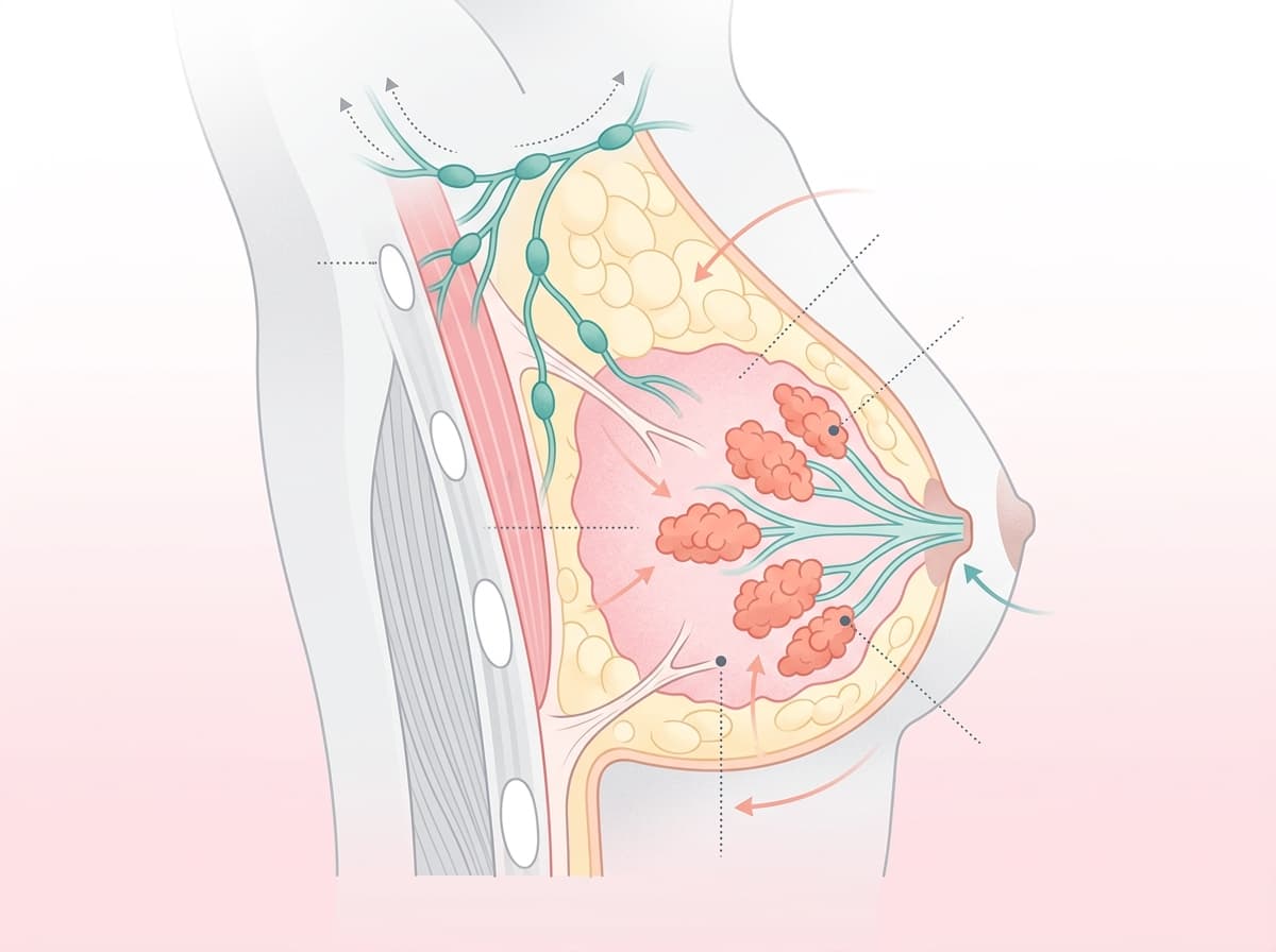
Not every breast lump is cancer. Approximately 80% of breast lumps are benign (non-cancerous) — including fibroadenomas, cysts, and fibrocystic changes. However, every new or unusual lump deserves a medical evaluation. Never ignore a change in your breast — early assessment is always the safest approach.
Types of Breast Cancer
Breast cancer is not a single disease — it is a group of cancers classified by where they start, how they grow, and their molecular receptor profile. Understanding the type is crucial because it directly determines the treatment plan, prognosis, and long-term management.
Types of Breast Cancer — Classification by Origin, Growth Pattern & Receptor Profile
Scroll table →| Type | Description | Frequency | Key Characteristics |
|---|---|---|---|
| Ductal Carcinoma In Situ (DCIS) | Non-invasive; cancer cells confined within the milk ducts | ~20–25% of new cases | Stage 0; highly curable; treated with surgery ± radiation |
| Invasive Ductal Carcinoma (IDC) | Cancer starts in the ducts and invades surrounding breast tissue | ~70–80% of all breast cancers | Most common type; can spread to lymph nodes and other organs |
| Invasive Lobular Carcinoma (ILC) | Cancer starts in the lobules and invades surrounding tissue | ~10–15% | Often harder to detect on mammography; may appear in both breasts |
| Triple-Negative Breast Cancer (TNBC) | Lacks ER, PR, and HER2 receptors | ~10–15% | More aggressive; does not respond to hormone or HER2-targeted therapy; treated with chemotherapy and immunotherapy |
| HER2-Positive Breast Cancer | Overexpresses HER2 protein, promoting rapid cell growth | ~15–20% | Historically aggressive but now has excellent outcomes with targeted therapies like trastuzumab (Herceptin) |
| Inflammatory Breast Cancer (IBC) | Rare, aggressive form; cancer cells block lymph vessels in breast skin | ~1–5% | Causes redness, swelling, warmth; often mistaken for infection; requires urgent treatment |
Staging: How Far Has the Cancer Spread?
Breast cancer staging uses the TNM system (Tumour size, Node involvement, Metastasis) and ranges from Stage 0 (non-invasive, in situ) to Stage IV (metastatic, spread to distant organs). Staging is critical for determining treatment approach and estimating prognosis.
Breast Cancer Staging — TNM Classification & 5-Year Survival Rates
Scroll table →| Stage | Description | 5-Year Survival Rate (Approx.) |
|---|---|---|
| Stage 0 (DCIS) | Non-invasive; cancer confined to the ducts | ~99% |
| Stage I | Small tumour (≤2 cm); no lymph node involvement | ~95% |
| Stage II | Tumour 2–5 cm and/or spread to 1–3 axillary lymph nodes | ~80–85% |
| Stage III | Locally advanced; larger tumour or extensive lymph node involvement | ~50–70% |
| Stage IV | Metastatic; cancer has spread to distant organs (bones, liver, lungs, brain) | ~20–30% (varies widely with treatment) |
9 Early Warning Signs of Breast Cancer
Early-stage breast cancer is often painless and invisible — which is precisely why awareness of subtle changes is so critical. Here are the 9 warning signs every woman (and man) should know:
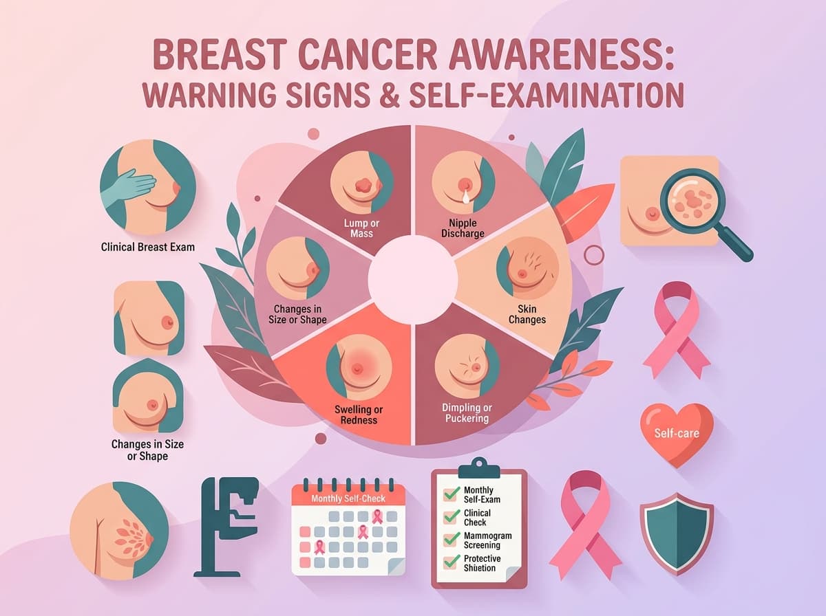
9 warning signs every woman should know:
- A new lump or thickening — A hard, painless, irregularly shaped lump in the breast or underarm (axilla) that feels different from the surrounding tissue. This is the most common first symptom.
- Change in breast size or shape — Unexplained asymmetry, swelling, or a noticeable change in the contour of one breast that doesn't follow your menstrual cycle pattern.
- Skin dimpling or puckering — When the skin over the breast pulls inward, creating a dimpled or 'orange peel' texture (called peau d'orange), it may indicate an underlying tumour pulling on the tissue.
- Nipple inversion or retraction — A nipple that suddenly turns inward (inverts) or changes direction, especially if this is a new development and affects only one breast.
- Nipple discharge — Spontaneous, unexplained discharge from one nipple — especially if it is clear, bloody, or straw-coloured — requires immediate evaluation. Milky discharge from both breasts is usually hormonal and benign.
- Redness, warmth, or swelling — Persistent redness, warmth, or swelling of the breast skin that resembles an infection but doesn't respond to antibiotics may indicate inflammatory breast cancer.
- Persistent breast pain — While most breast pain is hormonal and benign, localised pain in one spot that doesn't change with your menstrual cycle and persists for weeks should be evaluated.
- Skin changes around the nipple — Flaking, scaling, crusting, or ulceration of the nipple or areola skin (may indicate Paget's disease of the nipple, a rare form of breast cancer).
- Swollen lymph nodes — Hard, painless lumps in the armpit or above the collarbone may indicate that cancer cells have spread to the lymph nodes, even before a breast lump is noticeable.
Seek immediate medical attention if you notice: A rapidly growing lump, bloody nipple discharge, sudden nipple inversion, breast skin that looks red or inflamed like an orange peel, or a hard lump under your armpit. These could indicate aggressive forms of breast cancer that require urgent evaluation.
Causes & Risk Factors
Breast cancer develops due to a combination of genetic, hormonal, lifestyle, and environmental factors. While some risk factors are beyond your control, understanding them helps you make informed decisions about screening and prevention.
Key causes and risk factors for breast cancer:
- Gender — Being female is the single biggest risk factor. Women are about 100 times more likely to develop breast cancer than men, though male breast cancer does exist.
- Age — Risk increases with age. In India, the peak incidence occurs between 40–55 years, though younger women (30+) are increasingly being diagnosed.
- Family history & genetics — Having a first-degree relative (mother, sister, daughter) with breast cancer roughly doubles your risk. Inherited mutations in the BRCA1 and BRCA2 genes carry a 45–72% lifetime risk of breast cancer.
- Early menstruation or late menopause — Starting periods before age 12 or reaching menopause after age 55 increases lifetime oestrogen exposure, which is linked to higher breast cancer risk.
- Nulliparity or late first pregnancy — Women who have never been pregnant or who had their first child after age 30 have a slightly higher risk due to prolonged oestrogen exposure.
- Hormone replacement therapy (HRT) — Long-term use of combined oestrogen-progesterone HRT (for more than 5 years) modestly increases breast cancer risk.
- Obesity (especially post-menopausal) — Excess body fat increases oestrogen production after menopause, raising breast cancer risk. Indian women with a BMI >30 are at 1.5–2x higher risk.
- Physical inactivity — Sedentary lifestyles are linked to higher breast cancer incidence. Regular exercise (150+ minutes/week of moderate activity) reduces risk by 20–30%.
- Alcohol consumption — Even moderate alcohol intake (1 drink/day) increases breast cancer risk by about 7–10% compared to non-drinkers, with risk increasing with consumption.
- Dense breast tissue — Women with dense breasts (more glandular and fibrous tissue than fatty tissue) have a 1.5–2x higher risk and are harder to screen with mammography alone.
- Previous chest radiation — Women who received radiation therapy to the chest area (e.g., for Hodgkin's lymphoma) before age 30 have a significantly elevated risk.
- Previous breast conditions — Atypical ductal hyperplasia (ADH), atypical lobular hyperplasia (ALH), or lobular carcinoma in situ (LCIS) increase future breast cancer risk.
How to Do a Breast Self-Examination (BSE)
Breast self-examination (BSE) is a free, empowering tool that helps you become familiar with the normal look and feel of your breasts so you can quickly spot any changes. The ICMR recommends that all women above 20 years practise monthly BSE. Perform it 3–5 days after your period ends (when breasts are least swollen). Post-menopausal women should pick a fixed date each month (e.g., the 1st).
Step 1: Visual Inspection — Stand Before a Mirror
Stand in front of a mirror with shoulders straight and arms on your hips. Look for any changes in the size, shape, or symmetry of your breasts. Check for skin dimpling, puckering, bulging, redness, or rashes. Check if either nipple has changed position or become inverted. Repeat with arms raised overhead.
Step 2: Look for Nipple Discharge
Gently squeeze each nipple between your thumb and index finger. Check for any discharge — clear, milky, yellow, or bloody. One-sided bloody or clear discharge is especially important to report to your doctor.
Step 3: Feel Your Breasts While Lying Down
Lie down on your back (this spreads the breast tissue evenly). Use the pads of your three middle fingers (not the tips) of the opposite hand to examine each breast. Use small circular motions about the size of a ₹1 coin. Cover the entire breast from collarbone to bra line, and from armpit to cleavage. Use three levels of pressure — light (just under the skin), medium (mid-tissue), and firm (close to the ribcage).
Step 4: Feel Your Breasts While Standing or Showering
Many women find it easiest to feel their breasts when their skin is wet and slippery, such as during a shower. Repeat the same circular motion technique, covering the entire breast and armpit area. Check for any lumps, hard knots, or thickening that was not there before.
Step 5: Check Your Armpits
Do not forget to check the axillary (armpit) area. Breast tissue extends into the armpit, and the axillary lymph nodes are the first place breast cancer spreads. Feel for any hard, painless lumps or swelling in this area.
Pro Tip: BSE is NOT a replacement for clinical examination or mammography — it is a complementary awareness tool. If you notice ANY change during BSE, don't panic — 80% of lumps are benign — but always consult a doctor for proper evaluation. At Sanjivani Hospital, Virar, our surgeons offer rapid-access breast clinics for same-week assessment.
Screening Guidelines: When to Get a Mammogram
Screening aims to detect breast cancer before symptoms appear, when it is most treatable. The Indian Council of Medical Research (ICMR) and the Breast Imaging Society of India (BISI) recommend a combined approach of clinical breast examination (CBE) and mammography, adapted to the Indian context where breast density tends to be higher in younger women.
ICMR Breast Cancer Screening Guidelines by Age Group
Scroll table →| Age Group | Breast Self-Examination (BSE) | Clinical Breast Examination (CBE) | Mammography | Additional Notes |
|---|---|---|---|---|
| 20–29 years | Monthly | Every 3 years | Not routinely recommended | Ultrasound preferred for evaluating lumps in young women with dense breasts |
| 30–39 years | Monthly | Every 1–3 years | Only if high-risk (BRCA+, family history, prior chest radiation) | Baseline mammogram may be recommended around age 35 for high-risk women |
| 40–49 years | Monthly | Annually | Every 1–2 years | Annual mammography strongly recommended; ultrasound may supplement for dense breasts |
| 50+ years | Monthly | Annually | Every 1–2 years (biennial is acceptable) | Continue screening as long as life expectancy is 10+ years |
| High-Risk (any age ≥30) | Monthly | Every 6 months | Annually + Breast MRI annually | Includes BRCA1/2 carriers, prior chest radiation, Li-Fraumeni syndrome, strong family history |
Dense Breasts & Indian Women: Indian women tend to have denser breast tissue compared to Western populations, especially before age 40. Dense breast tissue can mask tumours on mammography. If your mammography report says you have "heterogeneously dense" or "extremely dense" breasts, ask your doctor about supplementary ultrasound or breast MRI screening.
Diagnosis: The Triple Assessment
If a suspicious lump or change is detected — either by you, during CBE, or on mammography — the gold-standard diagnostic approach in India (as recommended by ICMR) is the Triple Assessment:
The three components of the Triple Assessment:
- 1. Clinical Examination — A detailed physical examination of both breasts, armpits, and supraclavicular (above collarbone) areas by an experienced breast surgeon or surgical oncologist.
- 2. Imaging — Bilateral mammography and/or breast ultrasound (USG). MRI may be added for high-risk patients or to assess the extent of cancer before surgery.
- 3. Tissue Biopsy — The definitive step. A core needle biopsy (preferred over fine needle aspiration/FNAC) extracts a tissue sample for histopathological examination, grading, and receptor testing — ER (Oestrogen Receptor), PR (Progesterone Receptor), HER2/neu status, and Ki-67 proliferation index. These results determine the precise cancer subtype and guide personalised treatment.
When to See a Doctor
Consult a doctor immediately if you experience any of the following:
- Any new lump in the breast or armpit that persists for more than 2 weeks
- Nipple discharge that is spontaneous, one-sided, clear, or bloody
- Sudden nipple inversion or change in nipple direction
- Skin changes on the breast — dimpling, puckering, redness, rash, or 'orange peel' texture
- Persistent, localised breast pain that doesn't change with your menstrual cycle
- Change in breast size or shape that is new and unexplained
- A family history of breast or ovarian cancer — consult a doctor about genetic counselling and early screening even if you have no symptoms
- You are 40 years or older and have never had a mammogram
Medical & Surgical Treatment Options
Breast cancer treatment is highly personalised and depends on the type, stage, receptor status, and the patient's overall health. Modern treatment in India follows globally standardised protocols and is available at a fraction of the cost compared to Western countries.
Surgical Options
Surgical Treatment Options for Breast Cancer
Scroll table →| Procedure | How It Works | Best For | Recovery Time | Approximate Cost in India |
|---|---|---|---|---|
| Lumpectomy (Breast-Conserving Surgery) | Removes only the tumour and a margin of surrounding healthy tissue; breast shape is preserved | Stage I–II with small tumours; followed by radiation | 2–3 weeks | ₹1,00,000 – ₹2,50,000 |
| Modified Radical Mastectomy (MRM) | Complete removal of the breast tissue along with axillary lymph nodes | Stage II–III; large or multi-focal tumours; patient preference | 3–4 weeks | ₹1,50,000 – ₹4,50,000 |
| Sentinel Lymph Node Biopsy (SLNB) | Identifies and removes only the first 1–3 lymph nodes to which cancer would spread | Early-stage cancers; avoids unnecessary full axillary clearance | 1–2 weeks | Included with primary surgery |
| Breast Reconstruction | Rebuilds the breast shape using implants or autologous tissue (flap surgery) after mastectomy | Any patient desiring restoration after mastectomy; can be immediate or delayed | 4–6 weeks | ₹2,00,000 – ₹5,00,000 |
Systemic Therapies
Systemic Therapies for Breast Cancer — Chemotherapy, Radiation, Hormone, Targeted & Immunotherapy
Scroll table →| Therapy | How It Works | Used For | Duration | Approximate Cost Per Cycle |
|---|---|---|---|---|
| Chemotherapy | Cytotoxic drugs kill rapidly dividing cancer cells throughout the body | Adjuvant (post-surgery), neoadjuvant (pre-surgery to shrink tumours), or palliative | 4–8 cycles over 3–6 months | ₹20,000 – ₹1,00,000/cycle |
| Radiation Therapy | High-energy X-rays destroy microscopic cancer cells remaining after surgery | After lumpectomy (mandatory); after mastectomy for large tumours or node-positive disease | 5–6 weeks (daily sessions) | ₹1,00,000 – ₹3,50,000 total |
| Hormone (Endocrine) Therapy | Blocks oestrogen/progesterone from fuelling ER+/PR+ cancer growth (Tamoxifen, Letrozole, Anastrozole) | ER-positive and/or PR-positive breast cancers | 5–10 years (oral daily medication) | ₹500 – ₹3,000/month |
| Targeted Therapy (Anti-HER2) | Monoclonal antibodies (Trastuzumab/Herceptin) specifically target HER2 protein on cancer cells | HER2-positive breast cancers | 1 year (every 3 weeks) | ₹20,000 – ₹1,50,000/cycle (biosimilars significantly cheaper) |
| Immunotherapy | Boosts the body's immune system to recognise and attack cancer cells (Pembrolizumab) | Triple-negative breast cancer (TNBC) with PD-L1 expression | Varies; often combined with chemotherapy | ₹1,00,000 – ₹3,00,000/cycle |
Financial Support: The Ayushman Bharat (PM-JAY) scheme provides up to ₹5 lakh per year in cashless coverage for cancer treatment at empanelled hospitals. Additionally, organisations like the Cancer Patients Aid Association (CPAA) offer subsidised chemotherapy drugs. Many Indian pharma companies now manufacture affordable biosimilar versions of trastuzumab, bringing the cost of HER2-targeted therapy down by 60–80%.
Prevention: 10 Evidence-Based Steps to Lower Your Risk
Evidence-based strategies to reduce your breast cancer risk:
- Perform monthly breast self-examination (BSE) — Starting from age 20, examine your breasts 3–5 days after your period ends. Familiarity with your normal breast tissue is your first line of defence.
- Get regular mammograms — Follow ICMR screening guidelines: annual mammography from age 40 (or earlier if high-risk). Mammography can detect cancers up to 2 years before they become palpable.
- Maintain a healthy weight — Aim for a BMI between 18.5–24.9. Post-menopausal obesity significantly increases breast cancer risk due to excess oestrogen production in fat tissue.
- Exercise regularly — Engage in at least 150 minutes of moderate-intensity exercise per week (brisk walking, swimming, cycling, yoga). Exercise reduces circulating oestrogen and insulin levels, lowering cancer risk by 20–30%.
- Limit alcohol intake — If you drink, limit to no more than 1 drink per day. Even moderate alcohol increases breast cancer risk. Non-drinkers should not start drinking.
- Breastfeed if possible — Breastfeeding for 12+ months cumulatively over a lifetime reduces breast cancer risk by approximately 4.3% for every 12 months of nursing (Lancet Collaborative Group study).
- Eat an anti-inflammatory, plant-rich diet — Increase intake of cruciferous vegetables (broccoli, cauliflower, cabbage), berries, turmeric (haldi), green tea, flaxseeds, and omega-3 fatty acids. Reduce processed meat, refined sugars, and trans fats.
- Avoid unnecessary hormone therapy — If you need HRT for menopausal symptoms, use the lowest effective dose for the shortest duration. Discuss risks with your gynaecologist.
- Know your family history — If you have 2 or more first-degree relatives with breast or ovarian cancer, consult a genetic counsellor about BRCA1/BRCA2 testing. Early knowledge allows proactive screening and risk-reduction strategies.
- Avoid smoking and environmental toxins — While the link between smoking and breast cancer is modest, tobacco smoke contains carcinogens that damage DNA. Minimise exposure to pesticides, endocrine-disrupting chemicals, and industrial pollutants.
Myths vs Facts About Breast Cancer
Common Breast Cancer Myths Busted with Evidence-Based Facts
Scroll table →| Myth | Fact |
|---|---|
| Only women with a family history get breast cancer | 75–80% of women diagnosed with breast cancer have NO family history. Every woman is at risk, and screening is important for all. |
| Wearing an underwire bra causes breast cancer | There is no scientific evidence linking bras (of any type) to breast cancer. This is a persistent myth with zero basis in medical research. |
| Breast cancer always presents as a painful lump | Most early-stage breast cancers are painless. Pain is NOT a reliable early symptom. Many cancers are detected on mammography before any lump is felt. |
| Small-breasted women are less likely to get breast cancer | Breast size has no bearing on breast cancer risk. Risk depends on genetics, hormones, lifestyle, and tissue density — not cup size. |
| Mammograms cause breast cancer due to radiation | The radiation dose from a mammogram is extremely low (0.4 mSv — equivalent to 7 weeks of natural background radiation). The benefit of detecting cancer early far outweighs this minimal risk. |
| If I have a BRCA mutation, I will definitely get breast cancer | Having a BRCA1 or BRCA2 mutation increases risk to 45–72%, but it does NOT mean cancer is inevitable. Risk-reduction strategies (enhanced screening, preventive medication, and in some cases prophylactic surgery) are highly effective. |
Dietary Do's and Don'ts for Breast Health
While no single food can prevent or cure breast cancer, a consistently healthy diet can lower your risk and support recovery during and after treatment. Here are evidence-based dietary recommendations with Indian food examples:
Dietary Recommendations for Breast Health — Indian Food Guide
Scroll table →| Eat More (Do's) ✅ | Limit / Avoid (Don'ts) ❌ |
|---|---|
| Cruciferous vegetables — Broccoli, cauliflower (gobi), cabbage (patta gobi), radish (mooli). Contain sulforaphane, which has anti-cancer properties. | Processed and red meat — Sausages, salami, bacon, excessive mutton. Linked to increased cancer risk when consumed regularly. |
| Turmeric (Haldi) — Active compound curcumin has potent anti-inflammatory and anti-cancer properties. Add to dal, sabzi, and golden milk. | Refined sugar and maida — White bread, pastries, biscuits, sweetened beverages. High glycaemic foods spike insulin, which can promote cancer cell growth. |
| Flaxseeds (Alsi) — Rich in lignans and omega-3 fatty acids; shown to reduce oestrogen activity. Add 1–2 tbsp ground flaxseeds to roti dough, smoothies, or curd. | Alcohol — Even 1 drink/day increases breast cancer risk by 7–10%. Reduce or eliminate alcohol consumption entirely. |
| Green tea — Rich in epigallocatechin gallate (EGCG), a powerful antioxidant. Aim for 2–3 cups daily. | Trans fats and hydrogenated oils — Vanaspati ghee, deep-fried snacks (samosas, pakoras in reused oil), commercial bakery items. Promote inflammation. |
| Seasonal fruits — Papaya, amla (Indian gooseberry), pomegranate (anaar), guava, berries. Rich in vitamin C, antioxidants, and fibre. | Soy supplements (high-dose) — While moderate dietary soy (tofu, soy milk) is safe, high-dose isoflavone supplements should be avoided, especially for ER+ breast cancer. |
| Whole grains — Brown rice, jowar, bajra, ragi, oats. Provide sustained energy and fibre that helps regulate hormones. | Excessive dairy fat — Full-fat paneer, cream, butter in large quantities. Opt for low-fat dairy or plant-based alternatives. |
| Nuts and seeds — Walnuts (akhrot), almonds (badam), pumpkin seeds. Anti-inflammatory and rich in healthy fats. | Packaged/ultra-processed foods — Chips, instant noodles, ready-to-eat meals. Contain preservatives and additives linked to chronic inflammation. |
Recovery: What to Expect After Treatment
A week-by-week recovery timeline after breast cancer treatment:
- Week 1–2 (Post-Surgery): Expect mild to moderate pain, swelling, and restricted arm movement on the affected side. Drainage tubes may be in place for 7–14 days. Begin gentle arm exercises as advised by your surgeon.
- Week 3–4: Most patients resume light daily activities. Surgical wound healing continues. Pathology results and receptor testing guide the next steps (chemotherapy, radiation, or hormone therapy).
- Month 1–6 (Chemotherapy Period, if needed): Chemotherapy is typically given in 4–8 cycles, every 2–3 weeks. Common side effects include fatigue, nausea (manageable with modern anti-emetics), hair loss (temporary — hair regrows after treatment), and lowered immunity. Maintain nutrition and hydration.
- Month 2–3 (Radiation, if needed): Radiation is usually given daily (Monday–Friday) for 5–6 weeks after surgery or chemotherapy. Side effects include localised skin redness and fatigue — both temporary.
- Month 6–12: Gradual return to full physical activity. Emotional recovery is just as important as physical healing. Join a support group or seek counselling if needed.
- Year 1–5 (Follow-Up): Regular follow-up visits every 3–6 months, annual mammography, and hormone therapy (if prescribed for 5–10 years). Most recurrences occur in the first 5 years, making follow-up adherence critical.
- Beyond 5 Years: Annual mammography and clinical examination continue lifelong. 5-year survivors of early-stage breast cancer have a recurrence risk of less than 10%. Many women lead full, active, cancer-free lives for decades.
Special Considerations
Breast Cancer During Pregnancy
Breast cancer during pregnancy is rare (about 1 in 3,000 pregnancies) but poses unique challenges. Diagnosis is often delayed because breast changes during pregnancy (engorgement, tenderness) can mask cancer symptoms. Ultrasound is safe for evaluation during pregnancy. Certain chemotherapy regimens can be safely administered during the 2nd and 3rd trimesters (after the first trimester, when organ formation is complete). Radiation and hormone therapy are delayed until after delivery.
Breast Cancer in Men
Yes, men can get breast cancer — though it accounts for less than 1% of all breast cancers. Risk factors include BRCA2 mutations, Klinefelter syndrome, family history, obesity, and liver disease. Symptoms are similar: a painless lump near the nipple, nipple discharge, or skin changes. Because awareness is low among men, male breast cancer is often diagnosed at a later stage. Treatment follows similar protocols as female breast cancer.
Young Women (Under 40)
Breast cancer in women under 40 is less common but tends to be more aggressive (higher-grade, more likely to be triple-negative or HER2-positive). Standard mammography may miss tumours in young women due to denser breast tissue — ultrasound or MRI are better screening tools for this age group. If you have a strong family history or known genetic mutation, discuss enhanced screening with your doctor starting at age 25–30.
Sanjivani Hospital, Virar — Your Partner in Breast Health. Our Department of General Surgery offers comprehensive breast care, including clinical breast examination, ultrasound-guided biopsy, and surgical treatment for breast conditions. We support patients through every step — from diagnosis to recovery. Book a consultation today by calling +91-XXXX-XXXXXX or visiting us at Sanjivani Hospital, Virar West, Maharashtra.
The Bottom Line
Key takeaways from this breast cancer awareness guide:
- Early detection is the single most powerful weapon against breast cancer — Stage I breast cancer has a survival rate exceeding 95%. Don't wait for symptoms; get screened.
- Perform monthly breast self-examinations from age 20 — Know what's normal for your breasts so you can spot changes early.
- Follow ICMR screening guidelines — Start annual mammography from age 40 (earlier if you're high-risk). Dense breasts may need supplementary ultrasound or MRI.
- Breast cancer treatment in India is world-class and increasingly affordable — From biosimilar targeted therapies to government schemes like Ayushman Bharat, financial barriers to treatment are shrinking.
- Breast cancer is NOT a death sentence — With modern treatment, millions of women survive and thrive. Don't let fear, stigma, or misinformation delay your diagnosis. This October, take the pledge to check, screen, and share awareness.